Views: 0 Author: Site Editor Publish Time: 2026-06-05 Origin: Site


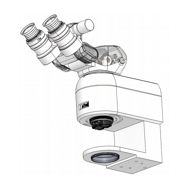
Ophthalmic surgery traditionally forces a hard divide in clinical practice. Surgeons plan complex procedures using static preoperative OCT scans. They then perform the surgery relying entirely on standard microscopic visualization. This legacy approach often leaves them flying blind regarding sub-surface tissue depth. Modern technology completely changes this dynamic. Today’s surgical platforms do not just work alongside Optical Coherence Tomography (OCT). They fully integrate MI-OCT into the optical path and Heads-Up Display (HUD).
This article serves as a comprehensive buyer’s evaluation guide. We know the clinical benefits of live volumetric imaging are well documented. However, clinical administrators and lead surgeons face complex procurement choices. You must evaluate hardware compatibility before buying. You need to assess workflow disruption and instrument shadowing risks. You also must calculate clinical ROI to justify the capital expense. We will explore how an integrated Ophthalmic Operating Microscope transforms the operating theater.
Integration is Reality: Modern systems provide live, 4D volumetric MI-OCT directly through the microscope oculars or HUDs, eliminating the need to pause surgery for handheld scanning.
Clinical Breadth: MI-OCT drives measurable outcomes in both anterior (e.g., verifying graft adherence in DSAEK) and posterior (e.g., visualizing macular hole peeling) segment surgeries.
Workflow Shifts: True integration offers "surgeon independence" via foot pedals and joysticks, removing reliance on imaging technicians.
Implementation Caveats: Buyers must account for instrument shadowing (metal vs. polycarbonate/silicone), potential surgeon cognitive overload, and the lack of dedicated MI-OCT reimbursement codes.
Ophthalmic imaging evolved rapidly over the last decade. Early intraoperative OCT required handheld external scanners. Surgeons had to halt the procedure entirely. They moved the microscope away. They brought the scanner over the sterile field. This process cost valuable operating room time. It also compromised focus and increased infection risks. Current Swept-Source MI-OCT (SS-MI-OCT) systems solve these problems natively. They build the OCT engine directly into the surgical viewing pathway.
Top-tier integrated systems deliver remarkable mechanical rendering capabilities. They generate up to 10 volumes per second. We call this 4D imaging, adding time to 3D spatial data. This speed enables a feature known as volume rotation. Surgeons can digitally rotate the live scan. They view the spatial relationship of pathology from multiple dynamic angles. You see the tissue architecture exactly as your instruments manipulate it.
Heads-Up Visualization (HUD) manages this data delivery. The system injects digital overlays directly into the oculars. It superimposes targeting boxes onto your surgical field view. These boxes show exactly where the scan coordinates sit. You also see real-time B-scan cross-sections floating above the tissue. This setup keeps your eyes on the patient. You never look away to check an external monitor.
Test the rendering latency during live demonstrations.
Check if the HUD brightness automatically adjusts to ambient room lighting.
Ensure the optical path shares the same focal plane as the surgical view.
Procuring an advanced Ophthalmic Operating Microscope requires clinical justification. The technology must demonstrate clear utility across multiple subspecialties. MI-OCT proves its value in both anterior and posterior segments.
Macular surgery benefits immensely from live subsurface visualization. Surgeons routinely peel the Internal Limiting Membrane (ILM). They typically rely on multiple dye applications to see this transparent layer. MI-OCT shows the membrane elevation in real-time. You can verify complete removal instantly. This reduces surgical dye toxicity risks.
High-risk pediatric cases highlight another critical advantage. Infants with Retinopathy of Prematurity (ROP) cannot cooperate for preoperative OCT. Surgeons often enter the eye without clear depth maps. MI-OCT maps the retinal traction precisely. Accurate depth measurement prevents iatrogenic retinal damage during complex dissections.
Corneal transplants rely heavily on precise tissue alignment. DSAEK and DMEK procedures demand perfect graft adherence. Surgeons previously used manual "S" stamps to verify graft orientation. MI-OCT eliminates this manual step. It immediately identifies interface fluid between the graft and the host cornea. You drain the fluid under direct OCT guidance. This dramatically reduces primary graft failure rates.
Cataract and glaucoma specialists also gain distinct advantages. They use MI-OCT to validate corneal incision architecture. They estimate phacoemulsification depth to prevent posterior capsule rupture. Glaucoma surgeons verify scleral flap thickness during trabeculectomy. They ensure precise implant positioning in MIGS procedures.
Clinical Applications of MI-OCT by Segment | ||
Segment | Procedure | MI-OCT Advantage |
|---|---|---|
Posterior | Macular Surgery | Real-time ILM peeling visualization; reduced dye usage. |
Posterior | Pediatric / ROP | Depth mapping without pre-op cooperation; prevents iatrogenic damage. |
Anterior | DSAEK / DMEK | Interface fluid detection; eliminates reliance on manual stamps. |
Anterior | Cataract | Incision architecture validation; phaco depth estimation. |
Technology should streamline the operating room. Legacy external scanners often did the opposite. They created severe workflow bottlenecks. You had to call a dedicated imaging technician. The technician would align the camera and adjust image parameters. This wait broke your concentration. Modern integrated platforms bypass this bottleneck entirely. They return control to the primary operator.
True integration demands hands-free control mechanisms. Surgeons achieve independence through automated and tactile UI solutions. You control the scanner via programmable foot pedals. You manipulate image rotation using joystick integration. Software provides automatic image optimization. You pull up volumetric imaging on demand. You maintain perfect sterility. You never break your surgical focus.
However, this autonomy introduces a new risk. We call it "information overload." You look into a single eyepiece. You see the analog surgical view. You see 2D cross-sectional scans. You process live 4D volumetric data. The human brain struggles to process all these data streams simultaneously. It can easily overwhelm a surgeon during critical maneuvers.
You must evaluate vendor software for user-centric design. The interface must be intuitive. It needs to allow rapid toggling of data layers. You should be able to turn off the 4D overlay with a single pedal tap. You want a clean surgical view when navigating complex bleeds. You bring the OCT data back only when verifying depth. A cluttered HUD causes fatigue. Software usability matters just as much as optical resolution.
Every imaging technology has physical limitations. You must transparently address these constraints before adoption. MI-OCT relies on light waves. Surgical instruments interact with these light waves. They cause optical attenuation and severe backscattering. This creates blind spots exactly where you need vision most.
Material compatibility dictates your success with MI-OCT. Standard instruments block the OCT beam. You must audit your surgical trays when procuring an integrated Ophthalmic Operating Microscope. Consider how different materials perform under the beam:
Metal Instruments: Titanium and stainless steel cause complete optical blockage. They project solid blackout shadows directly beneath the tool tip. You cannot see the underlying tissue.
Polyamide and Plastics: These materials cause moderate shadowing. They allow some signal through, but the image remains noisy and degraded.
Polycarbonate and Silicone: These are ideal materials for OCT-guided surgery. The infrared beam penetrates them easily. You clearly see both the instrument tip and the underlying retinal layers.
Fortunately, MI-OCT interacts well with typical surgical fluids. The OCT beam penetrates Brilliant Blue dye seamlessly. The signal remains strong even through dense staining. It also functions perfectly in air-filled cavities. Air-fluid exchanges do not disrupt the volumetric rendering. You maintain clear visualization throughout complex retinal detachments.
Using standard metal forceps during delicate OCT-guided peeling.
Failing to calibrate the z-axis focus after changing surgical fluids.
Ignoring the shadow angle. Tilt the instrument slightly to shift the blind spot away from the target zone.
Advanced imaging hardware represents a massive capital expenditure. Clinics routinely spend between $150,000 and $350,000 per unit. Generating a positive financial return proves challenging. The current medical billing landscape lacks dedicated reimbursement codes for intraoperative OCT. You cannot simply bill the patient for turning the scanner on. You must justify the investment through operational efficiency and clinical excellence.
We recommend building an ROI Justification Matrix. This framework helps hospital administrators see the broader financial picture. Direct revenue may lack, but indirect savings are substantial. Think about operating theater efficiency. Preventing a single primary graft failure in DSAEK saves thousands of dollars. Avoiding iatrogenic macular holes prevents costly secondary surgeries. You save non-billable theater time. You free up bed space. You reduce post-operative complication management.
ROI Justification Matrix for MI-OCT | ||
Value Pillar | Clinical Outcome | Financial Impact |
|---|---|---|
Secondary Surgery Reduction | Verified graft adherence; prevented macular holes. | Recoups non-billable OR time; reduces legal/malpractice risks. |
Surgical Efficiency | Faster decision-making; no waiting for technicians. | Increases daily case volume; optimizes staff allocation. |
Premium Positioning | Advanced capabilities for high-risk pediatric and complex cases. | Drives high-value referral networks; enhances institutional prestige. |
Your procurement strategy must include a rigorous training and rollout plan. New technology causes friction. Surgeons resist changing established routines. We recommend a phased adoption model. Do not start with complex retinal detachments. Start with straightforward macular cases. Let the surgical team master basic focusing and HUD navigation. Progress to dynamic image rotation later.
Use the integrated platform as a primary teaching tool. Fellows and residents benefit massively from seeing tissue depth live. The HUD allows the attending surgeon and the student to share the exact same volumetric perspective. This accelerates the learning curve for novice surgeons. It builds institutional expertise rapidly.
An ophthalmic operating microscope absolutely can, and should, integrate with OCT. This integration shifts the surgical paradigm. We move away from static preoperative planning. We embrace dynamic, real-time surgical response. Surgeons no longer guess tissue depth. They see it continuously.
Decision-makers must look beyond basic optical resolution when shortlisting vendors. You must evaluate software usability to prevent surgeon cognitive overload. You need robust foot-pedal autonomy. You must also secure a reliable supply of OCT-compatible surgical instrumentation. An amazing scanner fails if standard tools block its view.
Clinical directors should take immediate action. Request an in-theater demonstration. Do not accept a showroom walkthrough. Test the system during a live procedure. Focus specifically on the ease of UI toggling. Evaluate how the software handles instrument shadow management. See firsthand how surgeon independence transforms the operating room.
A: While some older models accepted external camera attachments, true volumetric MI-OCT with HUD overlays typically requires procuring a natively integrated system. These modern units are engineered with a shared optical pathway from the ground up.
A: It highly supplements visualization. It allows surgeons to verify membrane peeling in real-time. However, it does not entirely replace dyes. It usually reduces the need for repeated staining and lowers overall tissue toxicity.
A: Initial orientation takes only a few sessions. Mastering dynamic 4D image manipulation during complex maneuvers requires a phased approach. Surgeons typically begin with standard macular cases before advancing to complex detachments.

