Views: 0 Author: Site Editor Publish Time: 2026-06-08 Origin: Site


Upgrading surgical equipment represents a high-stakes clinical decision for any medical facility. It directly impacts your operating room throughput. It also significantly affects surgeon longevity and overall patient outcomes. Modern cataract surgery demands a flawless red reflex. You need precise depth perception and exceptional ergonomic sustainability to perform safely. Today, the medical equipment market is heavily saturated. Options range from traditional analog scopes to advanced 3D digital displays. This overwhelming variety complicates the procurement process immensely.
You need a structured, evidence-based evaluation framework to navigate this landscape. This framework must bridge technical specifications with practical clinical realities. You will learn how to balance optical clarity, illumination engineering, and biomechanical workflow. We will guide you through evaluating apachromatic lenses, coaxial illumination angles, and wireless foot pedal integrations. By understanding these core elements, you can confidently select the ideal system for your specific operating environment.
Base your procurement on the balance of optical clarity (apochromatic lenses, parfocality) and depth of field required for your specific cataract procedures.
Prioritize low-angle (0–2 degree) coaxial LED illumination to ensure a stable, resilient red reflex while minimizing retinal phototoxicity risks.
Factor in surgeon ergonomics—specifically working distance flexibility (200–400mm) and programmable wireless foot pedals—to reduce musculoskeletal fatigue and improve OR turnover.
Evaluate Total Cost of Ownership (TCO) beyond the initial purchase price, heavily weighting vendor calibration services, modularity for intraoperative OCT, and LED lifecycle.
You must establish clear procurement baselines before looking at technical specifications. Start by evaluating your surgical frequency and the primary user base. High-volume clinics typically host multiple surgeons rotating through the same operating room. They require systems offering highly programmable user profiles. You can preset specific magnification steps and diopter baselines for each doctor. This reduces inter-case setup times significantly. It keeps your surgical schedule running efficiently.
Next, consider your exact sub-specialty requirements. Pure anterior segment practices focus heavily on cataracts and MIGS procedures. They possess specific depth-of-field requirements. Practices combining anterior and posterior segment procedures face different challenges. They need versatile optical platforms. These platforms must handle rapid shifts between front-of-eye and back-of-eye visualization. You must align your hardware choices with your primary surgical load.
Space constraints and portability also dictate your options. Assess your operating room footprint carefully. Distinguish between fixed ceiling mounts, wall mounts, and heavy floor stands. Floor stands often feature robotic balancing for superior stability. However, they consume valuable floor space. Alternatively, you might require portable units. Portable units work well for multi-clinic deployment. They allow you to share high-end equipment across different physical locations.
Assess your daily surgical volume and set realistic OR turnover targets.
Map out the exact physical dimensions and structural limits of your operating room.
Identify all surgeons using the equipment to determine user profile requirements.
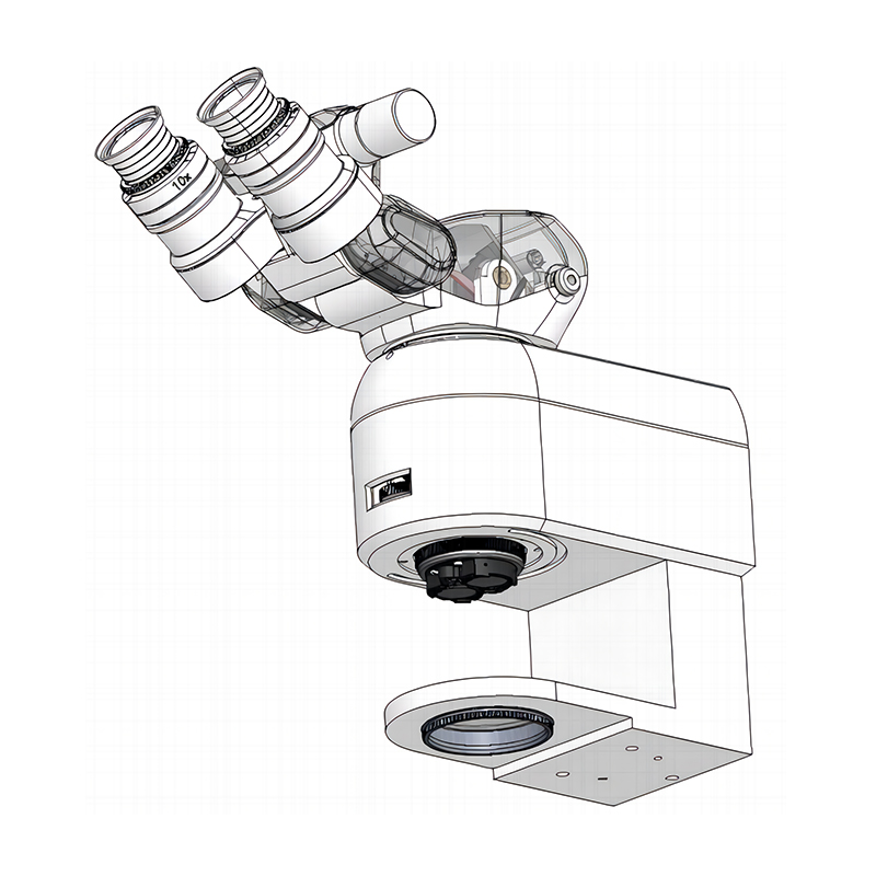
Optical clarity remains the foundation of safe cataract surgery. You must insist on genuine apochromatic optics. These specialized lenses correct chromatic aberrations across red, green, and blue wavelengths. This correction ensures true-to-life tissue rendering. It prevents frustrating color fringing around dense cataracts. Sharp tissue contrast helps you distinguish the anterior capsule during complex cases.
Verify the system's parfocal capabilities meticulously. Parfocality ensures the surgical field remains sharply in focus during rapid magnification shifts. You frequently transition from 16-20x magnification for capsulorhexis down to 8-10x for fragment removal. The system must retain absolute focus during these jumps. If it fails, you waste valuable time manually refocusing. This constant adjustment disrupts your surgical rhythm.
You must acknowledge a strict physical trade-off in optics. High magnification improves minute resolution but aggressively shrinks your depth of field. Deep spatial awareness relies on a generous DOF. Evaluate proprietary technologies designed to bypass this limitation. Some manufacturers fuse optical pathways intelligently. They combine one high-resolution beam in one eye and one high-DOF beam in the other. Your brain merges these images effortlessly. This reduces the need for constant manual refocusing during lens extraction.
Assess the stereo base width carefully. A 25mm stereo base provides optimal 3D depth perception. It gives you the spatial awareness required to maneuver instruments safely inside the anterior chamber.
Optical Visualization Trade-offs in Surgical Microscopes | |||
Optical Parameter | Primary Benefit | Clinical Limitation | Ideal Surgical Stage |
|---|---|---|---|
High Magnification (16x - 20x) | Exceptional detail and resolution. | Shallow depth of field; requires frequent refocusing. | Capsulorhexis, delicate tissue manipulation. |
Low Magnification (8x - 10x) | Broad spatial awareness and high DOF. | Reduced micro-detail visibility. | Nuclear fragment removal, IOL insertion. |
Fused Optical Pathways | Balances both resolution and DOF simultaneously. | Proprietary technology; limits vendor options. | Entire cataract procedure. |
Compare traditional binocular eyepieces against digital 4K/3D visualization systems. Digital heads-up systems unbind the surgeon from physical oculars. They drastically improve cervical spine ergonomics. You can sit upright while viewing a large external monitor. However, you must carefully evaluate adoption risks. Digital systems introduce a slight learning curve. You must adapt to minor screen latency. Digital tissue rendering looks different from pure optical light. You also need distinct OR lighting adjustments. Hybrid systems offer a practical fallback. They allow you to switch instantly between digital screens and analog eyepieces.
A stable red reflex is the core driver of safe cataract surgery. It illuminates the lens opacities against the bright retinal reflection. Evaluate scopes offering 0–2 degree small-angle coaxial illumination. This specific angle eliminates deep-cavity shadows effectively. It maintains the red reflex even if the patient's eye shifts slightly. When evaluating an Ophthalmic Operating Microscope, this illumination geometry determines your clinical success.
Compare collimated beams against strictly focused beams. Collimated beams offer a broader red reflex zone. They accommodate minor eye movements beautifully. Focused beams provide intense localized contrast instead. Many surgeons prefer collimated options for standard cases because they offer a wider margin of error during patient movement.
The transition from traditional light sources to LED technology is non-negotiable today. Traditional fiber-optic halogen or xenon sources draw approximately 180W. Direct LED illumination uses under 25W. This massive reduction in power consumption significantly reduces thermal tissue damage risks. It prevents accidental macular burns during prolonged cases. LED technology also offers a 50,000-hour operational lifespan. It eliminates mid-surgery bulb blowouts completely.
Ensure the illumination system strictly adheres to ISO 10936-2 standards. It must feature robust ultraviolet (UV) and infrared (IR) filtration. Unfiltered light causes severe retinal and corneal phototoxicity. Safety filters protect your patients while you operate.
The objective lens focal length dictates your physical working distance. This distance typically ranges from 200mm to 400mm. Your procurement choice must match the physical stature of your surgical team. A tall surgeon struggles with a short focal length. It also must accommodate the physical space required for instrument maneuvering above the patient's eye.
Assess ergonomic add-ons thoroughly. Inclinable binocular tubes are essential. They allow you to adjust the viewing angle independently of the microscope body. Optical extenders push the eyepieces closer to you. These features prevent "over-accommodation". They stop you from leaning forward awkwardly. Proper alignment prevents chronic neck and back strain over a long career.
Choose adjustable objective lenses offering variable ranges (e.g., 175mm to 225mm).
Invest in 0-180 degree inclinable binocular tubes for maximum positional flexibility.
Ensure the optical head maintains a low profile to prevent spatial conflicts.
Evaluate wireless, programmable foot switches carefully. Surgeons must control X-Y axis positioning effortlessly. They need to adjust zoom, focus, and illumination continuously. They must execute all these functions without breaking sterility or visual contact. Wireless pedals eliminate hazardous cables from the OR floor. They improve room safety immediately.
Investigate the physical mapping compatibility carefully. The microscope’s foot pedal must align with the phacoemulsification machine’s pedal. You typically control the microscope with your non-dominant foot. Your dominant foot manages fluidics and ultrasound energy. Seamless bilateral foot control is critical for surgical rhythm.
Your surgical equipment should have a reliable operational lifecycle of 7–10 years. You must evaluate the chassis's ability to accept future integrations. Medical technology evolves rapidly. You might want to add intraoperative OCT (optical coherence tomography) later. Digital astigmatism tracking overlays represent another highly valuable addition. The system architecture should support these upgrades seamlessly. You should never require a complete hardware replacement just to add a new software modality. Open architectures protect your initial capital allocation.
Consider long-term operational efficiency and mandatory downtime. Eliminating xenon bulb replacements with advanced LED technology improves daily reliability. Look for antimicrobial surface coatings on the chassis. These specialized coatings enable faster OR sterilization between cases. They improve your daily turnover rates substantially.
Scrutinize the vendor's Service Level Agreements (SLAs) closely. Precision optics require routine preventative maintenance to perform flawlessly. Dust accumulation and minor mechanical drifts occur naturally over time. Prioritize suppliers providing localized, certified technical support networks. Rapid local support prevents costly OR downtime. It ensures your surgical schedules remain entirely uninterrupted throughout the year.
Do not overbuy secondary digital features at the expense of core optical clarity.
Prioritize an unshakeable red reflex to ensure consistent patient safety during lens extraction.
Request comprehensive in-OR trial periods before signing final procurement documents.
Test ergonomics and foot pedal responsiveness under real-world surgical pressures.
Evaluate your visual fatigue levels during live cases; spec sheets cannot quantify comfort.
A: Typically, a working distance of 200mm serves as the industry standard. However, adjustable objective lenses allowing a range from 175mm to 225mm are highly recommended. This flexibility accommodates different surgeon heights, variable patient anatomies, and diverse surgical approaches without forcing doctors into awkward postures.
A: Coaxial lighting bounces light directly off the retina through the pupil. Specifically, a 0-2 degree angle of incidence is optimal. This tight angle creates the bright, glowing red reflex necessary to safely visualize the anterior capsule and assess dense lens opacities during delicate surgical maneuvers.
A: For high-volume clinics or teaching hospitals, the ergonomic benefits and shared screen visualization are often worth the required investment. They drastically reduce surgeon neck strain. However, solo practices must weigh the initial capital expenditure and necessary OR layout changes carefully against traditional, highly refined optical scopes.

