Views: 0 Author: Site Editor Publish Time: 2026-06-17 Origin: Site


Standardizing surgical equipment often seems like a smart strategy for hospitals. It simplifies inventory and streamlines daily routines. However, utilizing generic standard surgical microscopes for delicate eye surgeries causes significant problems. It compromises clinical outcomes. It also prolongs procedure times. General-purpose scopes simply lack the specialized features needed for ophthalmology.
An Ophthalmic Operating Microscope is engineered specifically for the unique anatomical constraints of the human eye. Eye procedures demand highly specialized illumination. They require seamless hands-free control. They also depend on zero-aberration optics. Standard multipurpose scopes cannot meet these strict requirements. Forcing a generic scope into an ophthalmic role places patient safety at risk.
This guide equips clinical directors, lead surgeons, and procurement teams with a clear comparison. We provide evidence-based insights to help you evaluate these systems. You will learn the core optical differences. You will discover ergonomic advantages. You will also see how digital integration boosts daily surgical volume. Ultimately, this clarifies why investing in a dedicated ophthalmic system improves your operational workflow.
Illumination is the primary differentiator: Ophthalmic scopes use Stereo Coaxial Illumination (SCI) to generate the crucial "Red Reflex" necessary for cataract surgeries—a feature standard scopes lack.
Hands-free operation is mandatory: Ophthalmic systems rely heavily on programmable, wireless foot pedals to manage zoom, focus, and lighting without breaking the sterile field or interrupting the surgeon's hands.
Digital integration drives modern ROI: Advanced ophthalmic models integrate intraoperative OCT and 3D heads-up displays (HUD), reducing surgeon fatigue and increasing daily surgical volume.
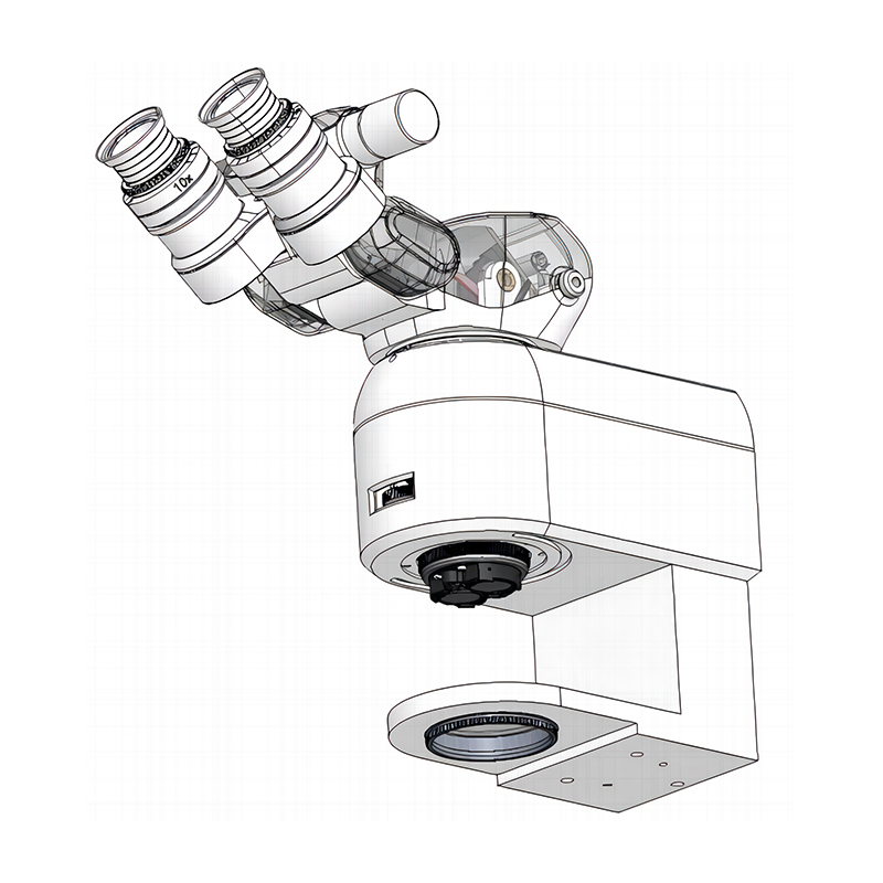
The differences between standard and ophthalmic scopes start at the foundational level of optics. General scopes prioritize deep cavity viewing. Ophthalmic scopes prioritize flat, transparent tissue visualization. This requires entirely different lighting techniques and lens structures.
Standard ENT or neurosurgery microscopes use offset lighting. This works well for deep tissue cavities. However, an ophthalmic operating microscope must align the illumination path almost parallel to the observation path. We call this Stereo Coaxial Illumination (SCI). This precise alignment generates a stable red reflex from the patient's retina.
The red reflex acts as a backlight. It provides vital contrast for procedures like capsulorhexis. The lens capsule is transparent. Without a strong red reflex, surgeons cannot see the capsule edges clearly. Standard scopes create shadows instead of backlighting. Shadows increase the risk of capsular tears. Such complications can cause the lens to drop into the vitreous. A dedicated ophthalmic scope prevents this by maintaining constant, brilliant coaxial illumination.
Working distance heavily influences surgical ergonomics and precision. Multipurpose standard scopes feature variable, longer working distances. They often range from 250mm to 300mm. This accommodates long surgical instruments used in spine or neurosurgery.
Ophthalmic procedures require a much shorter working distance. Surgeons sit very close to the patient's head. Objective lenses on eye scopes are strictly calibrated. They typically feature focal lengths of 150mm, 175mm, or 200mm. This optimized distance enhances hand-eye coordination. It gives you precise control over micro-instruments inside the anterior chamber.
Eye surgery demands absolute visual clarity. It requires zero chromatic aberration. It also demands zero spherical aberration. Color fringing around tissue edges causes dangerous miscalculations. High-end ophthalmic systems utilize fully apochromatic optics. These lenses bring all color wavelengths into a single focal plane.
Additionally, they feature specialized depth-management systems. The eye is a three-dimensional structure. You must see the cornea, lens, and retina clearly. Advanced depth-of-field technologies maintain a sharp focus on these intraocular structures. You avoid constant manual refocusing. This keeps the procedure moving swiftly and safely.
Feature | Standard Surgical Microscope | Ophthalmic Operating Microscope |
|---|---|---|
Illumination Type | Offset or angled lighting | Stereo Coaxial Illumination (SCI) |
Primary Advantage | Deep cavity shadow reduction | Stable red reflex generation |
Working Distance | Variable, long (250-300mm) | Fixed, short (150-200mm) |
Optical Focus | General tissue magnification | Apochromatic zero-aberration clarity |
Surgery is physically demanding. Ophthalmic surgeons often perform back-to-back procedures all day. Ergonomic strain ruins careers. Standard microscopes do not account for the rapid, micro-movements required in eye surgery. Ophthalmic systems solve this through intelligent design.
Ophthalmic surgeons use both hands continuously. One hand holds the phacoemulsification probe. The other hand manipulates a chopper or hook. You cannot reach up to adjust a microscope knob. Breaking the sterile field is strictly forbidden. It also breaks your concentration.
Dedicated ophthalmic scopes utilize multi-function wireless foot pedals. These pedals are highly programmable. They allow you to control X-Y positioning effortlessly. You can manage continuous zoom. You can adjust light intensity on the fly. The wireless architecture eliminates floor clutter. It prevents staff from tripping over thick cables during busy room turnovers.
Traditional oculars force surgeons into sustained neck flexion. You bend over the eyepieces for hours. This causes severe cervical spine strain. Many high-volume surgeons develop chronic back pain. Standard microscopes still rely heavily on this outdated physical posture.
Modern ophthalmic models replace traditional eyepieces entirely. They use 3D 4K, 55-inch digital displays. We call this a Heads-Up Display (HUD). You sit completely upright. You look straight ahead at the high-definition screen. This drastic ergonomic shift preserves postural health. It extends the career longevity of top surgeons. It also reduces daily physical fatigue dramatically.
A standard scope isolates the surgical view. Only the primary surgeon sees the intricate details. The rest of the operating room staff operates blindly. They must guess what the surgeon needs next based on verbal cues. This delays critical responses.
Ophthalmic systems with HUDs broadcast the surgical field to the entire room. This transforms the workflow. It brings incredible benefits to the whole team:
Scrub Nurses: They see the exact step of the procedure. They anticipate instrument hand-offs perfectly.
Anesthesiologists: They monitor patient micro-movements in real-time. They adjust sedation precisely when needed.
Surgical Assistants: They gain superior contextual awareness. They can manage unexpected complications proactively alongside the lead surgeon.
Trainees: Residents see exactly what the attending surgeon sees, in full 3D, accelerating their learning curve.
The modern eye clinic runs on digital data. Standard tissue scopes are analog tools. They magnify what is optically visible. They do nothing more. A dedicated ophthalmic system functions as a digital workflow hub. It processes live data to enhance surgical precision.
Optical Coherence Tomography (OCT) is vital for retinal specialists. Historically, OCT was only available as a preoperative clinic device. Premium ophthalmic microscopes now overlay real-time OCT scans directly into the surgeon’s view. This changes everything for posterior segment surgeries.
Consider macular hole repairs. Surgeons must peel extremely thin membranes from the retina. A standard scope only shows the tissue surface. An integrated OCT system shows depth. You see the distinct tissue layers in real-time cross-sections. You know exactly where your forceps are located. This prevents accidental damage to healthy retinal layers.
Ophthalmic units feature specialized digital filters. Standard scopes lack these algorithmic enhancements. For example, Minimally Invasive Glaucoma Surgery (MIGS) requires you to navigate the trabecular meshwork. This tissue has very low natural contrast. Digital filters highlight this specific anatomical structure instantly.
Furthermore, these filters allow you to operate under extremely low light levels. The retina is highly sensitive to intense light. Prolonged exposure causes macular phototoxicity. Digital enhancement amplifies the image computationally. You protect the patient's vision while maintaining perfect surgical clarity.
Modern ophthalmic microscopes sync seamlessly with clinic Electronic Medical Records (EMR). They pull preoperative biometry data directly into the operating room. This enables powerful Augmented Reality applications during cataract surgery.
Placing a Toric Intraocular Lens (IOL) requires perfect axis alignment. Traditionally, surgeons mark the patient's eye manually with an ink pen before surgery. Ink smears. Ink washes away. Manual marking introduces human error. Connected microscopes project the exact digital alignment axis onto the live surgical view. The system tracks the eye. It guides the IOL placement flawlessly. This eliminates manual marking errors and guarantees better refractive outcomes.
Hospital administrators often view standard equipment as a safe bet. However, examining clinical throughput reveals a different reality. Specialized ophthalmic scopes generate massive operational efficiencies. They eliminate bottlenecks that plague standard surgical setups.
Clinical data highlights the power of optimized illumination. Switching to direct LED ophthalmic scopes reduces average procedure times. Surgeons do not struggle with poor contrast. They do not pause to adjust lighting constantly. Studies show dedicated scopes can reduce average cataract surgery time by up to 2.6 minutes per procedure.
This time savings compounds quickly. High-volume Ambulatory Surgery Centers (ASCs) easily perform 10 to 15 procedures daily. Saving nearly 30 to 40 minutes a day is significant. It equates to adding an additional reimbursable surgery to the daily schedule. It also reduces overtime hours for the nursing staff. Efficiency drives operational success.
Equipment failure mid-surgery is a nightmare. Standard scopes often use halogen bulbs. These bulbs degrade quickly. They require frequent replacements, sometimes after just 50 hours of use. If a bulb blows during a delicate vitrectomy, the surgery halts. You lose precious time swapping light sources.
Standard scopes also utilize fragile fiber-optic cables. Constant bending breaks the internal glass fibers. This degrades light transmission over time. Modern ophthalmic systems utilize direct-illumination LEDs. These LEDs boast up to 50,000-hour lifespans. They feature internal cable routing. There are no exposed fiber optics to snap. This drastically reduces unexpected downtime. Your operating room maintains constant, reliable uptime.
Choosing the right microscope requires a pragmatic approach. You must align the hardware with your specific clinical volume. Use this step-by-step evaluation lens when assessing a new Ophthalmic Operating Microscope for your facility.
Volume & Case Mix: Assess the ratio of anterior segment surgeries versus posterior segment surgeries. Cataract and cornea cases prioritize superior red reflex and short working distances. Retina and vitrectomy cases demand integrated wide-angle viewing systems. They also require inversion prisms, like the Porro-Abbe system, to flip the inverted image upright. Ensure the scope matches your primary caseload.
Light Safety & Patient Comfort: Evaluate the system’s lower illumination threshold. Patient photophobia causes unwanted eye movement under local anesthesia. Macular light toxicity is a severe clinical risk. The microscope must function brilliantly at 10% to 15% illumination capacity. It must prevent toxicity while maintaining crisp visibility for the surgeon.
Scalability & Modularity: Medical technology evolves rapidly. You do not want a closed system. Ensure the baseline optical carrier supports future upgrades. Can you add an assistant scope later? Can you upgrade from analog eyepieces to a 3D digital HUD? Can you attach an advanced 4K recording module for teaching purposes? Scalability ensures long-term clinical relevance.
Facility Constraints: Examine your physical operating room layout. ASCs often feature smaller rooms with lower ceiling load limits. Check if the ceiling structure supports a heavy ceiling-mounted variant. If not, evaluate the floor-stand models. Look closely at the footprint size. Ensure the floor base does not obstruct the surgeon's chair or the anesthesiologist's equipment cart.
Standard surgical microscopes are undoubtedly versatile tools for general hospitals. However, they introduce unacceptable compromises in ophthalmic settings. They deliver poor red reflex. They lack digital data overlays. They also force surgeons into painful, unergonomic postures. Eye surgery is too delicate for a generalized approach.
High-volume facilities should rethink their equipment strategy. You must view dedicated ophthalmic scopes not merely as optical tools. You should view them as digital workflow hubs. They drive clinical precision. They protect patient retinas. They also protect the surgeon's cervical spine.
The operational benefits heavily outweigh the perceived convenience of standardizing equipment. Faster surgeries and less clinical downtime lead to better daily throughput. As a next step, consult with your clinical directors. Run an operational throughput analysis. Compare your current standard scopes' daily efficiency against the accelerated workflow of a dedicated digital ophthalmic system. The clinical advantages will become immediately clear.
A: No. Standard scopes lack the precise Stereo Coaxial Illumination (SCI) required for eye surgery. They cannot generate a consistent red reflex. Without this red reflex, visualizing the transparent lens capsule is nearly impossible, making critical steps like capsulorhexis highly dangerous for the patient.
A: LED systems typically offer up to 50,000 hours of stable color temperature and brightness. In stark contrast, standard halogen bulbs suffer from rapid brightness degradation. They usually require mid-case replacement every 50 to 100 hours, which interrupts surgical workflow and causes unexpected clinical downtime.
A: Yes. Digital sensors require significantly less light to generate a clear, highly magnified image. This allows surgeons to operate at much lower illumination levels. Reduced light intensity directly lowers the risk of retinal light toxicity during prolonged, complex posterior segment procedures.

