
Views: 0 Author: Site Editor Publish Time: 2026-11-06 Origin: Site


Visualization serves as the absolute foundation of successful minimally invasive surgery. A surgeon's precision relies entirely on the clarity and perspective of their operative field. This reality makes the selection of optical equipment a critical factor for clinical outcomes. The rigid endoscope acts as the primary visual tool in modern operating rooms, utilized daily across specialties ranging from orthopedics and gynecology to general surgery.
However, these sophisticated optical instruments are far from one-size-fits-all. One of the most impactful procurement and operational decisions involves selecting the correct direction of view. Choosing an improper angle creates dangerous blind spots during a procedure. Conversely, selecting the optimal perspective ensures superior visualization, safer tissue manipulation, and accelerated patient recovery.
This engineering and clinical guide assists medical procurement teams, clinic managers, and surgeons in making highly informed decisions. By exploring the exact mechanics of clinical optics, you will learn how to match the right instrument to specific procedural demands. Furthermore, you will understand how modern manufacturing standards and digital sensor integrations maximize surgical efficacy.
Direction Dictates Navigation: The specific optical tilt, such as 0°, 30°, 45°, or 70°, determines the surgeon's line of sight. This directly impacts their ability to navigate around complex anatomical obstacles.
Straight vs. Angled Optics: A 0-degree scope offers a straightforward, intuitive view ideal for direct approaches. In contrast, angled lenses unlock crucial peripheral and lateral visualization for deep anatomical cavities.
The Versatile Standard: A 30-degree optical tilt remains the undisputed gold standard for abdominal procedures. It provides the perfect balance of forward trajectory and downward visibility.
Resolution is Paramount: Pairing an angled lens with high-resolution digital sensors maximizes clinical effectiveness, delivering distortion-free visuals across the entire surgical field.
Material Quality Matters: Purchasing decisions must heavily weigh rigorous optical testing, sapphire glass integration, and cross-brand compatibility to ensure reliable performance in demanding clinical environments.
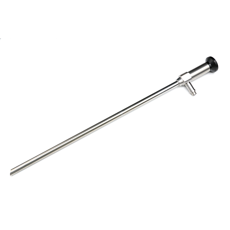
To make an optimal equipment selection, buyers must first understand the internal architecture of these delicate devices. The core of a standard clinical scope relies on a highly sophisticated Hopkins rod-lens system. This internal structure utilizes a series of precision-machined glass rods, separated by minimal air spaces, to transmit light and high-resolution images from the distal tip inside the patient back to the proximal eyepiece.
The Viewing Angle, technically referred to as the Direction of View, is a precise geometric measurement calculated relative to the mechanical longitudinal axis of the metal shaft. If the optical lens at the tip looks perfectly straight ahead, parallel to the shaft, engineers measure it as 0 degrees. If the internal distal prisms are engineered to tilt the view slightly downward or upward from that central axis, the angle of that tilt defines the instrument's perspective. This mathematical calibration dictates exactly where the center of the surgical image will focus when inserted through a trocar.
A frequent point of confusion among clinic managers and junior medical staff is the distinction between Field of View and the direction of view. While both metrics are vital for surgical visualization, they measure entirely different optical properties. Buyers must evaluate both specifications simultaneously when sourcing medical equipment.
The Field of View refers to the total width or cone of the observable image. A wider cone allows the surgeon to see a larger area of tissue at one time, functioning much like a wide-angle camera lens. The viewing direction, as established, refers to the directional tilt of that specific cone. For instance, a scope might feature a generous 80-degree Field of View but a 0-degree tilt. This means it captures a wide image directly in front of the tip. Understanding this distinction ensures the chosen instrument aligns flawlessly with the anatomical requirements of the intended surgery.
The 0-degree configuration provides a direct line of sight. Because the optical axis aligns perfectly with the mechanical axis of the instrument, the image on the surgical monitor corresponds exactly to the physical movement of the surgeon's hands. Moving the instrument left causes the anatomy on the screen to shift left in real-time.
Clinically, this straightforward perspective serves as the primary choice for procedures where the anatomical pathway is linear. Routine diagnostic cystoscopies, basic knee arthroscopies, and hysteroscopies rely heavily on this intuitive setup. Its primary limitation lies in its inability to look behind anatomical folds. If a dense tissue mass obstructs the direct line of sight, a 0-degree lens cannot visualize the hidden area without physically levering the entire instrument, which risks patient trauma.
In modern minimally invasive surgery, the 30 Degree angle is universally recognized as the most versatile option available. By incorporating a precise tilt at the distal prism, the scope allows surgeons to look downward, upward, or to the side, depending entirely on how the instrument is oriented along its axis.
The true engineering value of this angle is unlocked through rotation. By keeping the camera head stationary and physically rotating the light post of the scope, the surgeon sweeps the surgical field. This conical sweeping motion allows the surgical team to look over bulky organs or under overlapping tissue layers without applying dangerous torque to the patient's entry wound.
Steeper optical tilts are engineered for highly specialized clinical scenarios where extreme lateral or retrograde vision is absolutely mandatory. These are precision instruments designed for complex anatomical challenges rather than general-purpose use.
A 45-degree scope is frequently utilized in complex orthopedic joint explorations where navigating around dense bone structures is required. Meanwhile, a 70-degree angle serves as a critical tool in urology, specifically for retrograde viewing of the bladder neck. It is also essential in Ear, Nose, and Throat procedures where looking down at the vocal cords from a horizontal approach is necessary. The 90-degree scope provides true lateral vision, used in specific neurosurgical and sinus procedures. These extreme angles require advanced spatial awareness, as the visual orientation on the monitor can feel disconnected from the physical movement of the surgeon's hands.
Laparoscopy presents unique visual challenges. The abdominal cavity is densely packed with layered, overlapping organs. During these procedures, the abdomen is inflated with carbon dioxide to create a working space. In this dynamic environment, a 30-degree Laparoscope, typically in a standard 10mm diameter, is widely considered the gold standard.
Because entry ports are fixed in the abdominal wall, a surgeon has limited physical mobility. A straight 0-degree scope would require the operator to push the instrument deeply and at severe angles to see underlying structures. This risks trauma to the abdominal wall or internal organs like the liver. The 30-degree tilt solves this elegantly. It allows the metal shaft to remain relatively parallel to the patient's body while the angled lens looks down into the pelvic cavity or up toward the diaphragm, providing safe, comprehensive visualization.
Recent shifts in the medical device sector highlight a growing demand for advanced optical systems. As healthcare facilities globally transition toward complex, multi-quadrant minimally invasive procedures, including robotic-assisted surgeries, the reliance on versatile angled optics has surged. Standard straight scopes often fall short in providing the spatial awareness required for advanced tissue dissection. Consequently, hospitals are standardizing around 30-degree optics to reduce inventory complexity while accommodating higher surgical volumes.
Minimally invasive surgery fundamentally alters a surgeon's visual environment by projecting a three-dimensional anatomical space onto a two-dimensional monitor. Employing an angled lens actively mitigates this loss of depth perception through motion parallax. When the surgeon rotates the light cable, the angled lens sweeps across the cavity. Foreground anatomical structures move across the monitor faster than background structures, providing the surgeon's brain with critical spatial cues essential for safe tissue triangulation.
The physical construction of an angled lens requires the integration of microscopic prisms at the distal tip to bend incoming light. While highly effective for navigation, this optical bending can introduce minor visual anomalies, such as slight edge distortion or a marginal drop-off in light intensity at the periphery.
The integration of 4K Imaging technology has revolutionized how engineers manage these optical challenges. A 4K camera system provides four times the pixel density of standard High-Definition systems. This immense resolution, combined with advanced digital signal processing, effectively neutralizes edge distortion. It ensures that the extreme edges of a 30-degree view remain exceptionally sharp, allowing surgeons to identify micro-vascular structures with absolute confidence.
Medical procurement teams must recognize that a high-resolution digital camera head is only as effective as the optical glass it is attached to. Upgrading a surgical suite to a 4K system while utilizing older or poorly manufactured scopes creates a severe optical bottleneck. The ultra-sensitive sensor will simply magnify the optical flaws of the inferior lens, resulting in a pixelated image that negates the financial investment.
When sourcing equipment, administrators must ensure that the optical lenses are specifically engineered to resolve 4K signals. Premium models utilize precision-machined Hopkins rod lenses and flawless scratch-resistant sapphire distal windows. Ensuring this level of manufacturing guarantees that the entire imaging chain works in perfect harmony.
To streamline the procurement process, the following table breaks down the practical applications and engineering differences between the most common optical angles.
Viewing Angle | Primary Application | Depth Perception | Learning Curve | Best Suited For |
|---|---|---|---|---|
0-Degree | Direct, straight-line approaches | Low (relies on camera movement) | Minimal (Highly intuitive) | Routine diagnostic cystoscopy, basic arthroscopy |
30-Degree | Multi-quadrant sweeping and lateral views | High (utilizes motion parallax) | Moderate (requires rotational skills) | Laparoscopic cholecystectomy, complex gynecology |
70-Degree | Retrograde and extreme lateral visualization | High | Steep (orientation differs from hand movement) | Urology bladder neck viewing, specialized ENT |
The lifetime value and clinical safety of a medical scope depend entirely on its manufacturing standards. Medical facilities must prioritize instruments that undergo rigorous optical quality testing. A poorly constructed lens will degrade rapidly under the harsh conditions of a modern operating room.
High-quality Rigid Endoscope models utilize industrial-grade sapphire glass for the distal and proximal windows. Sapphire is exceptionally scratch-resistant, ensuring that the viewing angle is not compromised by accidental contact with other surgical instruments, such as shavers or graspers. Furthermore, the internal glass rod lenses must be securely mounted within a durable stainless steel body, laser-welded rather than glued, to withstand repetitive high-temperature sterilization.
A critical factor in the procurement process is ensuring seamless compatibility between the new scope and the hospital's existing surgical tower infrastructure. Angled lenses naturally require more powerful illumination than straight lenses because the distal prism absorbs a microscopic fraction of the transmitted light.
Therefore, buyers must ensure the scope is fully compatible with high-output LED or Xenon light sources. The instrument should feature universal light post adapters, accommodating standard fittings including ACMI, Wolf, and Storz. This universal compatibility prevents the need for costly secondary purchases of proprietary light cables and ensures the optimal viewing angle is always supported by adequate brightness.
The sophisticated internal optics that make an angled perspective possible are inherently delicate. The rod-lens system consists of fragile glass pillars suspended within a thin stainless-steel tube. Because of the precise alignment required at the distal prism, these instruments are highly sensitive to mechanical shock.
Mishandling the instrument, such as dropping it on a hard floor or using the shaft as a lever to retract dense tissue, can instantly shatter the internal glass rods. When this occurs, the viewing angle is destroyed, resulting in a half-moon shadow, a blurry field, or a completely black image on the surgical monitor.
To maintain the pristine clarity of the lens over hundreds of surgical procedures, strict adherence to sterilization protocols is required. While premium scopes are designed to be fully autoclavable at 134 degrees Celsius, the rapid temperature fluctuations inherent in steam sterilization place immense physical stress on the optical seals.
Surgical processing departments must ensure that the scopes are allowed to cool naturally. Artificially accelerating the cooling process by submerging a hot scope in cold sterile water causes micro-fractures in the optical seals. If these seals fail, moisture penetrates the shaft. During the next surgery, the heat from the light cable will vaporize this trapped moisture, fogging the angled lens from the inside and blinding the surgeon at a critical moment.
Selecting between a 0°, 30°, or 70° perspective depends entirely on the specific anatomical challenges of the surgical application. While straight lenses offer intuitive navigation for basic diagnostics, the 30-degree configuration stands out as the most versatile and indispensable choice for complex, multi-quadrant procedures.
Ultimately, combining the correct optical tilt with cutting-edge 4K resolution and rigorous manufacturing quality ensures surgical success, enhanced depth perception, and superior patient safety. Investing wisely in durable, high-transmission optics elevates the standard of care across any medical facility.
To find the perfect optical solution tailored to your surgical suite's exact specifications, explore precision-tested instruments by reviewing the robust product catalog at Rising-EO. Matching your clinical needs with their advanced engineering ensures long-term reliability and exceptional visual clarity.
While a 30-degree scope is highly versatile and utilized in the vast majority of abdominal surgeries, it is not universally mandatory. Some surgeons prefer a 0-degree scope for simpler, direct-approach procedures due to its easier spatial orientation and shorter learning curve. However, for complex cases requiring lateral views, the 30-degree angle is heavily preferred.
Yes, steeper angles like 70° or 90° experience a slight reduction in light transmission compared to a 0° scope due to the internal prisms required to bend the light. This is why pairing angled scopes with high-quality, high-intensity light sources and sensitive digital imaging systems is essential for maintaining optimal visibility.
Most modern scopes feature standard light post adapters and standard eyepieces, making them widely compatible across different brands. However, it is always recommended to verify compatibility prior to purchase, especially when upgrading to 4K cameras, to ensure the optical train can fully support the higher digital resolution.
Signs of optical degradation include a consistently blurry image, dark spots around the edges of your viewing angle, a yellowish tint to the tissue on the monitor, or persistent fogging after sterilization. Regular optical testing against a resolution target can help identify degradation before it impacts surgical performance.
In practice, a 0-degree laparoscope only looks straight ahead, meaning you must move the entire instrument to change your view. A 30-degree laparoscope looks slightly downward. By simply rotating the light cable without moving the shaft, the surgeon can achieve a sweeping, panoramic view of the abdominal cavity, allowing them to see behind organs and anatomical structures.
The choice of diameter depends on the surgical site and the patient. A 10mm diameter is the standard for adult laparoscopy, providing maximum light transmission and image resolution. Smaller diameters, such as 5mm or 2.9mm, are selected for pediatric cases, delicate joint arthroscopies, or procedures requiring smaller incisions, though they transmit slightly less light than their 10mm counterparts.


